MS205 – Abdominal Ultrasound
£447.00 (+VAT)
12 months access to recordings and course materials in included. Please note that these are webinar recordings and not live events. Full details on how to access the Mini Series will be emailed to you.
- Join Anna Newitt BVSc DVDI DipECVDI MRCVS for three 2-hour online sessions
- This Mini Series is suitable for all vets in practice, and will be of particular interest to those wishing to improve their confidence in Abdominal Ultrasound.
- Comprehensive notes to downloaded
- Self-assessment quizzes to ‘release’ your 8 hours CPD certification (don’t worry, you can take them more than once if you don’t quite hit the mark first time)
- A whole year’s access to recorded sessions for reviewing key points
- Superb value for money – learn without travelling
- Watch the recordings on any device
Programme
Session 1:
Liver, Spleen and Peritoneum
What you’ll learn:
- How do I position the patient for the best images?
- Patient preparation
- What will and what won’t damage your probe?
- Why does the liver look ‘small’ to inexperienced scanners?
- How to see as much of the liver and spleen as possible
- The importance of pressure
- Key structures to look out for
- Practical tips on assessment of liver size
- Recognizing free abdominal fluid and where to look for it
- How to find and scan the spleen
- Lymph nodes and masses
Urinary Tract and Adrenals
What you’ll learn:
- Best positioning for scanning the kidneys?
- Practical tips for getting the best view of the right and left kidneys
- Adapting your scan to body conformation
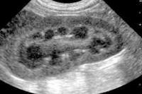
- How to differentiate renal cortex from medulla
- The appearance of normal kidney and common renal abnormalities
- Using the kidneys to find other organs
- Practical tips and common pitfalls in scanning the urinary bladder
- Recognizing stones, sediment, cystitis and tumours
- Finding the adrenals
Gastrointestinal Tract and Pancreas
What you’ll learn:
- Patient preparation for getting the best GI images
- Learn the distinctive appearance of the intestinal layers
- How to find and identify the stomach
- Explore the GIT with an ‘intestinal sweep’
- Specific characteristics of the colon
- Appearance of strictures and GI obstructions
- Landmarks for finding and imaging the pancreas
- Changes seen in pancreatitis for diagnosis and monitoring
- Practical advice on fine needle sampling
The price includes all 3 sessions, notes and quiz – 8 hours of CPD
*No traffic jams, accommodation hassles, pet or childcare, rota clashes, locum fees ……….. just great CPD and a valuable ongoing resource.
Course Feedback :
“I really liked how the speaker utilised real life videos of her scanning a live animal to illustrate where to put your probe when scanning for different organ systems. This was a really helpful tool to help with your visualisation of where to place the probe, how much pressure to use and what direction to angle it towards.”
“Given me a good basic knowledge and confidence to start doing ultrasound”
“The content of the Mini Series has Increased confidence finding structures and less obvious abnormalities”


